Glycolysis to Remember

Glycolysis to Remember

This video was very informative and understandable. The points discussed were clear and the lay out of the diagrams and structures shown were easy to follow. In all it was a very in depth lecture. Here are just some key points to remember about glycolysis pathway.

Glycolysis is mainly the splitting of glucose (6 carbons) into 2 molecules of pyruvate which each has 3 carbons. There are 10 enzymes reactions. 3 are irreversible while 7 are reversible.

The 10 enzymes are hexokinase, phosphohexoseisomerase, phosphofructokinase-1, aldolase, triosephophatisomerase, glyceraldehyde 3-phosphate dehydrogenase, phosphoglycerate kinase, phosphoglycerate mutase, enolase and pyruvate kinase.  

2 ATP and 2 NAD+ are consumed while 4 ATP and 2 NADH are mad. Hence the net gain is 2 ATP and 2 NADH.  

Every cell has glucose transporters where glucose can enter or leave the cell freely. By phosphorylating the glucose it essentially traps glucose into the cell. Since glucose-6-phosphate has no transporters and thus cannot leave the cell.

Triose phosphate isomerase is described as a typically kinetically perfect enzyme.

All kinase enzymes require Mg2+as their cofactor since it stabilises the charge from the ATP molecule.

The oxidative reaction produces energy needed to add the inorganic phosphor molecule to glyceraldehyde-3-phosphate.

For glycolysis to continue NAD+ needs to be converted to NADH and H+. Thus, NAD+ I limited in the cell.

Cell can make ATP via oxidative phosphorylation in glycolysis.

1, 3-bisphosphoglycerate is a very high energy molecule which adds a phosphate group to ADP molecule to make ATP molecule.  

Phosphoenol pyruvate has a lot of energy which is lost as heat.  

Carbohydrate video lecture

Carbohydrate video lecture

This video was based on many aspects of carbohydrates but mainly the polysaccharide forms.  Some information gained was that sucrose, lactose and maltose were most common disaccharides while starch, glycogen and cellulose were examples of polysaccharides. Glycosidic bonds link sugars together in disaccharides and polysaccharides. Ribose sugars are found mostly in RNA and DNA.

There are two types of glycosidic bond, N-glycosidic bond which links purines and pyridines in nucleotides and O-glycosidic bond which link sugars to one another in disaccharides. Glycosidic bonds are based on the position of carbon 1 hydroxyl group.

Hemiacetal is a cyclised sugar. When OH is blow in the pyranose ring structure it is a and when it is above it is b. A reaction of hemiacetal and an alcohol produces a polysaccharide that is an acetal.

It was highlighted that there is no enzyme that can breakdown cellobiose due to its linkage structure (chair formation).

Polysaccharides are polymers of the monosaccharide are used for storage and structural purposes. They can be branched or unbranched.

It was stated that starch and glycogen both have a (1→4) linkages but I will differ as only starch has this type of linkage. However, the video lecture did not mention that amylopectin was a form of starch which is similar in linkage to glycogen ( a (1→6) linkage). Cellulose has b (1→4) linkage and it is not branched and neighbouring chains are linked together by hydrogen bonds. A similar polysaccharide to cellulose is chitin.

There are glycoproteins which are proteins that were covalently linked to a carbohydrate unit by glycosidic bonds. In addition carbohydrates can form amino sugars.

This video lecture did not have points flowing in an easy manner and some points were confusing. However I was able to learn something from the video and enhance my understanding of the topic. An improvement would be to slow down the pace of the lecture so that points can be made clearer in certain cases such as the hemiacetal part. 

Uncoupling Protein 1 and Brown Adipose Tissue

Oelkrug, Rebecca, Maria Kutschke, Carola W. Meyer, Gerhard Heldmaier, and Martin Jastroch. 2010. “Uncoupling Protein 1 Decreases Superoxide Production in Brown Adipose Tissue Mitochondria.” The Journal of Biological Chemistry 285(29): 21961–21968. Accessed April 10, 2013. http://www.ncbi.nlm.nih.gov/pmc/articles/PMC2903373/  


Uncoupling protein 1 (UCP1) is used to catalyse the degeneracy of mitochondrial proton motive force in thermogenic brown adipose tissue and produce heat. The Brown Adipose Tissue (BAT) has a high oxidative capacity in which mitochondrial uncoupling could reduce lethal reactive oxygen species. BAT has a non-shivering thermogenesis which produces endogenic heat in mammals for instance. This helps to maintain a constant body temperature. Furthermore, the production of heat can, also, be activated by diets high in calories.

A high presence of mitochondria is a characteristic of brown adipose tissue. These mitochondria contain only a small amount of ATP synthase to convert nutrients into cellular energy. However, these mitochondria have a specialised uncoupling protein 1 which generates a proton leak in the inner membrane. This then, degenerates the proton motive force as heat.

            When BAT is exposed to the cold, lipolysis is stimulated and UCP1 is thus activated. Prolonged cold acclimation causes an increase in thermogenic capacity of brown adipose tissue due to enhanced UCP1 expression and recruitment of oxidative capacity. This then increases the chance of oxidative damage.

Electrons escape complexes 1 and 2 of the respiratory chain and this is the major source of cellular superoxide and the oxidative species that are then derived. Cold stress causes the activation brown adipose tissue mitochondria which can hence increase the influx of electrons. Furthermore, an increase in the probability of electrons to leak from the respiratory chai to form superoxide is prevented by UCP1 during b-oxidation which is the major pathway of substrate oxidation in BAT during cold induced thermogenesis. It was shown that UCP1 effectively lowers the production of mitochondrial superoxide during cold acclimation and fatty acid oxidation. Thus, oxidative stress can be reduced by uncoupling.     

I found this article a bit long and difficult to decipher much information. I was able to gain a small amount of information that furthered my knowledge on uncoupling protein 1 and brown adipose tissue. What a superoxide is and its prevention from forming was new information to me. The various experiments to test the hypotheses were not easy to follow but I think that I was able to get the gist of the article. Thus the importance brown adipose tissue and uncoupling protein 1 was noted.

A Look at Lactase Deficiency

 Kuokkanen, Mikko, Jorma Kokkonen, Nabil Sabri Enattah, Tero Ylisaukko-oja, Hanna Komu, Teppo Varilo, Leena Peltonen, Erkki Savilahti, and Irma Järvelä. 2006. “Mutations in the Translated Region of the Lactase Gene (LCT) Underlie Congenital Lactase Deficiency.” The American Journal of Human Genetics 78 (2): 339-344. Accessed April 13, 2013.  http://www.sciencedirect.com.ezproxygateway.sastudents.uwi.tt:2048/science/article/pii/S0002929707623647?np=y  


Congenital lactase deficiency (CLD) can be defined as a rare and severe gastrointestinal disorder. The main symptom is watery diarrhoea and occurs mainly in infants being breast fed or introduced to lactose containing formulas. The lactase gene is denoted by LCT. There are five distinguishing mutations that cause congenital lactase deficiency. The mutations alter the structure of the protein inactivating enzyme.  

As an autosomal, congenital lactase deficiency is recessively inherited. This results in decrease in lactase activity in intestinal mucosa and thus profuse watery diarrhoea is produced immediately after consumption of milk by the infant. This is owing to the fact that the mutations of the LCT gene cause the inability of the gene to activate the absorption of lactose when consumed. Infants with a lactose free diet results in the symptoms of watery diarrhoea, dehydration and weight loss being reduced.   

Of the five mutations LCT gene, three initiate premature shortening of lactase (FINmajor) and the other two cause the substitutions of amino acids of histidine for glutamine and serine for an uncharged glycine.  These mutations alter the structure of the polypeptide.

However, adult type hypolactasia is the opposite of congenital lactase deficiency as it is not rare but commonly found in adults. It is due to the development of down-regulation of lactase activity. A decline in lactase activity is quite normal but most European adults have lactase persistence as they retain lactase activity. Therefore, both of the lactase deficiencies are caused by DNA variants that affect the LCT gene.  

This article only got my interest as I wanted to know if congenital lactase deficiency was related to lactose intolerance. In a way it did to some extent as the symptoms were close (diarrhoea). However, this disorder was only seen in the early stages of infancy during breast feeding. I did not particularly like this article as the genetic information was not understandable. In the end I was still able to learn about a new disorder.     

Multiple Choice questions for Exams

Single Answer
1. In which of the processes is ATP made via the enzyme, phosphoglycerate kinase?
A. Substrate level phosphorylation
B. Oxidative phosphorylation
C. Photophosphorylation
D. Oxidative decarboxylation

2. Which statement about an allosteric enzyme is true?
A. It has one active site.
B. It has more than one active site and forms a sigmoid curve.
C. It has more than one active site and forms a hyperbolic curve.
D. It has one active site and forms a sigmoid curve.

Multiple Answers

A. 1 and 2 are correct
B. 2 and 4 are correct
C. 1, 2 and 3 are correct
D. Only 2 is correct
E. All are correct
3. Glycolysis is required for energy in which of the cells?
1) Muscle cells
2) Erythrocytes
3) Cells of the eyes
4) Cells of the brain

4. Km and Vmax are reduced by the same amount for
1) Mixed inhibition
2) Uncompetitive inhibition
3) Competitive inhibition
4) Non-Competitive inhibition

Enzyme Workout

So today I had a very big surprise – a pop quiz. My answers showed that I clearly was not prepared for it. The hardest thing was that I couldn’t remember which Lineweaver-Burk plot was for non-competitive inhibition. I think it’s very difficult to remember each one which as each has the same axes but different positions of the lines involved as well as where Vmax and Km are located.

Another problem was remembering the characteristics of the plots for the different types of inhibition as well as describing them. I had so much problems in this area that I had no answer for it. Lost most of my marks there far even more for having the wrong plot in the first place. Need to make a greater effort at this topic. 

Ionizable functional groups, pKa and pI

I think the process of calculating the titration of amino acids is fairly straight forward. The first structure must be fully protonated form. The equilibrium equations for each of the ionizations must then occur in increasing pKa values. This shows that there is a loss of acidity with an increase in pKa value.

After this the net charge is then calculated by adding together all of the charges present for each structure. Say there was COO, COO, NH3+ in the structure it would be an overall charge (2-) + (1+) = 1- . Hence the net charge of the ionized structure is 1-.

Finally, the pI is calculated. That is the isoelectric point where the pH at which the amino acid has a net charge of zero (0). It is calculated by finding the average of the two pK values for the structures immediately on either side of the this amino acid.

It’s not really difficult and I hope to succeed at this question for exams.

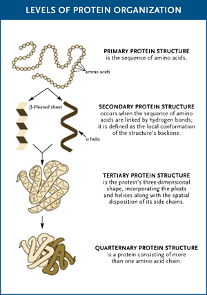
Missing a Few Things

Missing a Few Things

I just thought that it was a decent representation of folding as you advanced in the structure of the proteins. However, I thought that it missed the fact that the number of peptide chains increased with each new form of the protein structure.